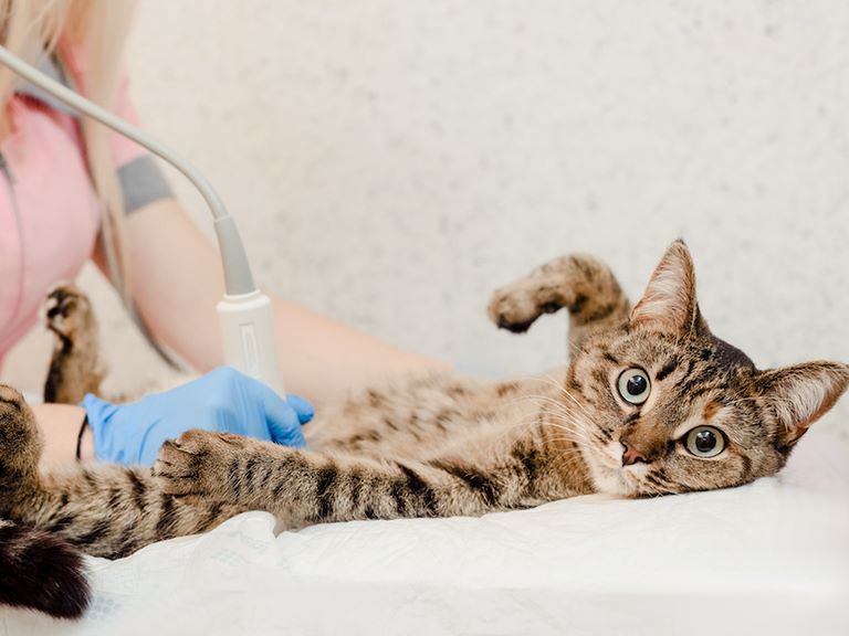
Ultrasound
What is an Ultrasound?
Ultrasonography is a non-invasive tool that uses sound waves to evaluate a body cavity (chest or abdomen), as well as to determine if there is any inappropriate fluid in that space. Unlike an x-ray that shows shadows of the organs, an ultrasound can assess the internal texture. For this reason, it is often more sensitive than an x-ray at picking up internal diseases, such as soft tissue changes in the GI tract, spleen, and liver. It is the only way we can assess lymph nodes within the abdomen. It also has the ability to more accurately assess anything abnormal detected on palpation by the doctor (is the firm mass an organ, a tumor, a lymph node, etc) . Sometimes ultrasonography gives a clear diagnosis, and sometimes it identifies an area that is abnormal to obtain tissue samples for more specific information or guides further testing.
Full abdominal ultrasounds help provide the best interpretation of what could be causing vague symptoms or lab work abnormalities that can be influenced by many different things. One example is liver enzyme elevations. These lab work changes can be caused by primary problems within the liver, but they could also be influenced by the health of the gallbladder, pancreas, or adrenal glands. As such, a full scan helps us understand the bigger picture, rather than having tunnel vision and focusing on only one organ.
We offer several levels of ultrasounds at Shiloh Animal Hospital
A focal scan is intended to look at one single organ or organ system (e.g. bladder, liver, spleen, etc). It is often used to investigate a specific symptom, such as blood in the urine. We also use this to recheck an abnormality observed on a previous ultrasound for response to treatment or changes to a lesion over time. This is interpreted in real-time by our doctors. It does not usually require sedation or admission to the hospital.
A brief or FAST scan broadens the scope of the ultrasound to a few key areas of the body. It may be used to assess the abdomen or chest for evidence of fluid (which can be caused by many different disease processes). For a brief abdominal scan, key points are imaged to look for evidence of disease in the main organs. At Shiloh we also offer this as a preventative measure for screening purposes as patients age, similar to the way routine lab work helps to track internal health. This scan may or may not require sedation or admission depending on the day and circumstance.
For a higher level of detail and sensitivity, a more complete abdominal ultrasound can be performed, imaging each organ in the abdomen. The imaging may be performed by our trained doctors and submitted for review by a specialist, or a radiologist may be scheduled to perform and interpret the scan in real time. This complete abdominal ultrasound is often performed under sedation to allow the patient to fully relax the abdominal muscles and to achieve best results; we will only sedate a pet if the owner has given permission to do so. The abdomen is fully shaved for this level of scan. The report is usually available within 48 hours.
Echocardiograms are ultrasounds of the heart. These are only performed by specialists that visit our hospital.
